
A novel “molecular flashlight” technology lets scientists study brain abnormalities without harming them. This boosts neurology research. The gadget can reach the brain without injury, making it somewhat intrusive. A very narrow light beam is sent out. Light reveals nerve tissue’s chemical nature. It detects molecular changes caused by tumours or other harm. This “molecular flashlight” is now used for research, but specialists believe it could one-day aid patients. Results are published in Nature Methods.
Brain molecular changes connected to cancer and neurological disorders have been difficult to explore without invasive interventions. A new ultra-thin tool delivers light into mice’s brains for molecular analysis. Today’s Nature Methods article (December 31) incorporates international collaboration, including the Spanish National Cancer Research Centre (CNIO) and the Spanish National Research Council. Researchers call this finding a “molecular flashlight” since it shows nerve tissue’s chemistry. This approach helps scientists detect molecular changes in primary and metastatic brain cancers and TBI.
Molecular Spotlight for Brain
One-micron probes under 1 mm are molecular spotlights. Human hairs are 30–50 microns, therefore they may be injected safely into the brain. The flashlight probe may be useful for monitoring molecular changes induced by severe brain damage and diagnosing brain metastases, but it is not yet suitable for human trials. The European NanoBright partnership, led by Manuel Valiente of the CNIO’s Brain Metastasis Group and Liset Menéndez de la Prida of the CSIC’s Neuronal Circuits Laboratory, performed the research. Biomedical research Italian and French scientists founded NanoBright, where they conducted biomedical research.
Molecular Flashlight Technology Breakthrough
Light can record or start brain activity, which is pretty cool. However, this idea has been around for a fairly long time. Light is one example of something that can be used to control what a single cell does. This is what we refer to as optogenetics. However, cells need to have a gene added that makes them sensitive to light for these ways to work. NanoBright’s new technology lets scientists study the brain without changing it first. This is a big step forward for research in the area. Vibrational spectroscopy is what the new molecule flashlight is based on.
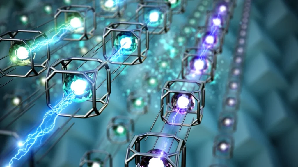
Light scatters differently on molecules depending on their chemical composition and shape. We capitalize on this. We can locate each molecule’s signal or band that way. The range indicates tissue type based on chemicals. This technique “lets us study the brain in its natural state, without changing it first,” explains Manuel Valiente. This technique also lets us analyze any brain area, including genetic markers or abnormalities. Vibrational spectroscopy shows alterations in brain chemicals during illness.
Neurosurgery already uses Raman spectroscopy, albeit less accurate and more invasive. Studies have examined its application during brain tumour surgery, according to Valiente. After thetumour’ss main part is surgically removed, a Raman spectroscopy probe can detect any remaining cancer cells.” However, this is only possible if the brain is open. The hole is sufficiently large. We cannot use these relatively large “molecular flashlights” in live animal models without causing them significant harm.
Minimally Invasive Brain Analysis
A word for “minimally invasive.” NanoBright probes hardly damage brain tissue. Authors endorse Nature Methods.” It said Valiente’s CNI group discharged tumor-front cells without surgery in brain metastasis models using molecular spotlights. “The difference with existing technology is that we can now perform this analysis in a minimally invasive way, regardless of whether the tumour is superficial or deep.” The CNIO team says the probe may “differentiate various oncological entities, such as types of metastases, based on their mutational profiles, by their primary origin, or from different types of brain tumours.”
AI in Brain Diagnostics
The Cajal Institute studied epileptogenic zones surrounding traumatic brain injuries. Epileptic seizure-prone brain areas exhibit distinct vibrational signatures due to tumour or damage. Menéndez de la Prida asserts that different locations impact molecular fingerprints differently and suggests using AI to diagnose diseased organisms. “We will be able to find new high-precision diagnostic markers by combining vibrational spectroscopy with other ways of recording brain activity and advanced computer analysis that uses artificial intelligence,” the CSIC person says. “This will facilitate the development of advanced neurotechnology for new biomedical applications.”
Related: AI Monetization Software Growth Drives Industry Profit in 2025
Summary
Brain researchers have developed a “molecular flashlight” to observe brain biochemical changes without invasiveness. This new technique illuminates the brain with ultra-thin probes to reveal nerve tissue’s chemical composition. The “molecular spotlight approach ” detects brain tumours, metastases, and TBI-induced molecular changes.
International teams from the Spanish National Cancer Research Centre (CNIO), CSIC, and NanoBright collaborated on the Nature Methods study. Vibrational spectroscopy lets scientists study brain activity and chemical changes without surgery or genetic manipulation. This finding might transform brain diagnoses and therapy by providing a less invasive technique to diagnose brain cancers, epilepsy, and brain metastases.
Despite promising results, the technology is not ready for human testing. It may enable high-precision diagnostics and improved neurotechnology for biomedical applications. Adding AI to this strategy might improve its ability to find novel brain illness indicators.